In this section, the research and
projects are shared...
(Only principles and ideas are explained in brief since they are IP, please bear with me!)
________________________________________________________________________________


I
am basically an Electrical Engineer but my research area is in Biomedical
Engineering. My specialized focus is on Imaging, Instrumentation and diagnostic
instrumentation; but certainly it is not boundary for my research!
I
have worked on
- Optical imaging for medical
diagnosis,
- Diabetic foot ulcers,
- Photo-acoustic imaging &
spectroscopy
- Microwave imaging
- and so on...
Few
projects on which I worked previously are briefly explained below.
1) Near infrared imaging and
spectroscopy:
In
this technique, light in the range of 700-1100 nm wavelength from an
electromagnetic spectrum is used as source of energy to shine the biological
tissue of interest and reflected or transmitted or backscattered light from the
same tissue is detected from the sensors. After the signal conditioning, the
output is analysed qualitatively. There are several challenges, governing
parameters, selection of suitable instrumentation and understanding the theory
of the mechanism in the tissue. All these need to studied and addressed.
 |
| Near Infrared Imaging |
 |
| Near Infrared Spectroscopy |
This method is cost
effective and has potential to characterize the tissue; also to detect
anomalies present in the tissue such as tumor/haematoma. Therefore, near
infrared imaging & spectroscopy is emerging as new diagnostic tool in
medical field.
2) Time correlated single photon counting for optical
spectroscopy and imaging applications:
This is based on optical technique for the
detection of photons in time domain. The laser light is detected from a photodector
and the signal is very weak. Meaning that, only few photons out of input light
are able to detect from the tissue. Therefore, a non-linear crystal based on
SHG (Second Harmonic generation) is used for counting the few output photons
through time correlation analysis. The system is not only cost-effective but
also highly sensitive. Its main application is in the detection of breast
tumors.
(A*STAR Sponsored Project, NUS, Singapore)
3) Photo-acoustic imaging and spectroscopy:
Optical imaging such as diffuse optical
imaging has major drawback of light scattering through the tissue due to which
the resolution is poor. When the photons penetrate in to the tissue the light
energy is absorbed by the tissue cells. The tissue cells gained energy then
propagates in to the surrounding in the form of acoustic waves. So, these
acoustic waves are detected from ultrasound sensors from the surface of the
tissue. Light energy is used as in input source but the detected output in the
form of acoustic waves, hence this technique is called as photo-acoustic
method. The input light source may be single wavelength or multi-wavelength
laser source emitting in form of short pulses.
An experimental setup of the PA system is
shown in below picture (Courtesy; Pittsburgh University, USA).

4) Microwave imaging system for biomedical applications:
Microwave imaging is a promising technique
in which electrical property distribution in the human body are mapped for
medical applications. It has been widely researched specifically for early
stage detection of breast cancer. Recently, this imaging technique is widely
researched because of its high contrast, patient comfort and safety at low
cost. An UWB patch antenna transmits the microwave frequency propagating
through the target and another the receiving antenna receives the backscatterd
microwave signal in the medical band of frequency (2.4 and 5.8 GHz). The signal
is measured and analysed through a network analyser. The data is stored in
computer and then plotted for electrical parameters to reconstruct image of the
object. The patch antenna for transmitter and receiver is designed, simulated
and fabricated then used for medical imaging applications.
5) Medical image segmentation:
Routine clinical examination may not be
enough for any conclusive diagnosis of the disease in several occasions.
Imaging plays an important role for the clinical diagnostic and therapeutic
procedures. The radiographic imaging varies according to their pathologic
features. Image segmentation refers to partitioning of an image into
distinct regions of interest that contain pixels with similar attributes. The
main goal of segmentation is to differentiate the object of interest from the
background of image. Thus the segmentation of image may be considered as an important
step in image processing techniques. Accurate segmentation of medical images
becomes a key step in image processing which can help for suitable planning of
diagnosis and treatment of the patient. This technique can guide the
radiologists precisely for a specific diagnosis of lesions or delineating
pathological regions, thus limiting the differential diagnosis. Several
algorithms do the segmentation on images but Watershed method, which is based
on natural phenomenon of water flow in lakes, is attracting large number of
researchers due to its fast action, simple algorithm to implement in computer
and versatile for twin images too. The morphological watershed image
segmentation is implemented on MRI images to extract calvarial bone (skullcap)
from the scans. It is useful for evaluation of calvarial lesions, calvarial
bone grafts and calvarial bone fractured patients that could help in diagnosis
and treatments. The same algorithm is extended to segment the cranial
vault bone from radiologic images of human head.
6) Early detection of Alzheimer's disease:
Alzheimer’s Disease (AD) is related to
human brain which affects a significant fraction of the aged people causing
problems with short-term memory, thinking, spatial orientation and behavior and
worsening over a period of 20 years ultimately leading to death of the patient.
Currently, there is no cure for the disease. Although the onset of AD cannot
yet to be stopped or reversed, an early detection and diagnosis could be
helpful to the people with AD a better chance to benefit from available drug
and non-drug therapies that may improve their health condition and enhance
their quality of life. Electroencephalography (EEG) is considered as one
of the most important tests for the diagnosis of neurological and
neurophysiological disorders which is a measure of the electrical activity of
the brain. When the neurons of the brain are activated they produce electrical
signal. Neurons are mutually connected in the form of closed neuronal networks.
If there is any discontinuity in the path of the network leading to the damage
of anatomical and physiological tracking of the circuitry would result in
steady decline of cognition that will be an early indication of Alzheimer’s.
The study is in progress. The below schematic shows the proposed method of
recording the brainwave data.
From this study the
probable biomarkers of AD are investigated. The structure of the study is shown
in the below figure.
This method is
non-invasive, painless, cost-effective and can be repeated several time for
thorough investigation to diagnose the disease in its early stage.
7) Diagnosis of Parkinson's disease from
SPECT images:
Parkinsonism (PS) is a clinical syndrome described
by tremor, bradykinesia, rigidity, and postural instability. The name
“parkinsonism” is derived from the progressive neurodegenerative illness called
Parkinson’s disease (PD). Although there could be a number of underlying
causes, PD remains the most common cause of parkinsonism. The accuracy of the
clinical diagnosis of PD is still limited as there is no standard diagnostic
test is available. Furthermore the disease is incurable; however a pre-stage
diagnosis can help to minimize the progression of the disease. Though
there is no specific clinical test to confirm the PD, but imaging of brain is
considered as an effective diagnostic procedure since it gives visual insight
of the brain in the form of an image. Traditional diagnostic imaging with CT or
MRI brain scans does not show changes in the brain but they give anatomical
pictures of the brain. To identify the disease in early stages, often
functional imaging is preferred than the diagnostic imaging. For the
definitive diagnosis of the PD, specialized imaging techniques are required for
histologic examination of Intraneuronal Lewy body inclusion of substantia nigra
compacta, which are not possible practically in life. However, the studies
suggest that the nigrostriatal projection loss is characterized with the PD
symptom which is associated with the striated dopamine deficiency.
Clinically, two technologies are commonly
used as nuclear imaging and they are; positron emission tomography (PET) and
single photon emission computed tomography (SPECT). They provide the means for
detecting in-vivo metabolic and neurochemical changes of PD. These imaging
technologies made possible to visualize functional, chemical processes and
metabolic activities in the targets by using radiotracers as agents. SPECT imaging
is widely accepted clinically to conform the loss of dopamine chemical and this
type of scan is known as dopamine transporter scan or DaTSCAN, by making
possible to use as an aid that can help diagnosing the PD in early
stages. Thorough investigation of the scans could able to detect dopamine
transporters. However, for reasonably correct assessment of the images,
appropriate algorithms are essential to play the role in the diagnosis of the
disease more accurately. In our on-going work, we aim to classify the
stage of the PD so that medical expert can know at which stage the PD is
present in the patient. In this study, we to use sparse learning techniques to
develop some novel classifiers by utilizing the power of Neural Networks.
8) Human gait evaluation system for
rehabilitation of elderly people:
Human gait refers to locomotion achieved through the movement of
human limbs. Since the gait is a largely automated motor task it becomes an
identity of a person’s style and quality of life. Often, gait can be used as a
biometric measure to recognize known persons and classify unknown subjects.
Visual inspection of the person’s gait also indicates the health condition of
the subject.
Observation of gait is an important aspect of diagnosis that may
provide information about several musculoskeletal and neurological conditions.
Reliable cognition of gait characteristics over time, continuous monitoring,
evaluation and proper analysis have demonstrated their significance in
clinical, sports, rehabilitation, personnel training and robotic research. In
the field of biomedical engineering, gait analysis and evaluation is attracting
the researchers.
Gait is altered by different factors, such
as age, gender, working conditions, body weight, etc. Ageing has great
impact on gait and its pattern changes drastically with normal ageing. It is
therefore necessary to understand the kinematic and kinetic parameters of gait
pattern and hence to take preventive steps against abnormalities related to
gait disorders. In this project, it is proposed to observe the gait pattern
from normal healthy people and lower limb affected aged people. It is also
proposing to suggest suitable corrective measures for improvement in
abnormalities. The sketch shows experimental setup to acquire gait pattern
data from three directions.
There are about 25 gait
abnormalities. The types of abnormalities while examining the patients are
demonstrated in the below video.
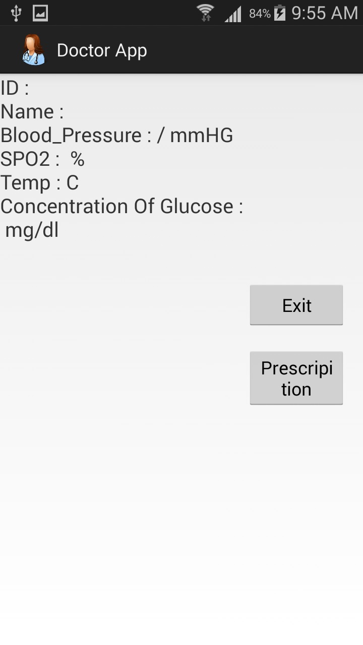
9) Mobile phone app for bio-telemetry:
Rapid advancements in communication
technology have spread to medicine also. Particularly, smartphone technology
has made medical provisioning through mobile systems a reality. Innovations in
mobile software application are potential benefits to the public health since
the mobile platforms became more user-friendly, computationally powerful and
are affordable. The innovative mobile apps can contribute in clinical
consultation complementing face-to-face interaction in the health care at lower
risk to the public. We have developed and evaluated mobile app for smartphone
on Android platform to facilitate interaction between the patient and doctor
where the patient seeks advice, diagnosis and treatment from the doctor from
remote places. The Graphic User Interface (GUI) display screens of the
smartphones are incorporated the medical data needed by the clinician to
interpret and respond to information. The scheme of the project and
screen-shots from mobile phone are shown below.


The medical application
software for quick diagnosis helps the patients to get treatment plans remotely
by saving the time of travel to visit the clinic. The doctors can diagnose and
treat the patients by this app at their convenient time from their places. Thus
the time and cost of both patients and doctors will be saved. The software is
user-friendly without involving many operations and no computers or dedicated
software are required. It is an open source, freely accessible software to
benefit the user whoever need this app. Installation of the app in the
smartphone is quite simple and more useful to socio-economically poor people as
well as rural dwelling patients.
10) Electrosurgery Unit:
Electrosurgery unit is a helpful tool in all areas of the surgical
field from the most basic surgery as wart removal, or hair removal to the most
sophisticated surgeries as open heart, orthopedic, and transplant
procedures. The main principle of this device is the conversion of
electricity into heat.
Also, it is capable of producing a cutting and/or coagulating
clinical effect on tissue by the use of alternating current at a high
frequency. Voltages and Currents can be changing depending on the desired
clinical effect. The idea tried in this project helps to make surgery in
short time, decrease the cutting and dangerous coagulation, minimize the loss
of blood in surgery and cancellation of the use of sharp surgical tools. The
block diagram of the project is shown below.
The unit developed is
only prototype and is tested clinically. However, the design features and the
product would allow for real tests only standardization following regulatory
norms.
No comments:
Post a Comment